Compared With Rods Cones Are Quizlet
Learn vocabulary terms and more with flashcards games and other study tools. Which statement is true about scotopic vision as compared to photopic vision quizlet.

Eye Micro Anatomy Of Retina Anatomy The Retina Notes
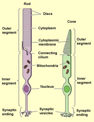
Rods and cones are the two types of photoreceptor cells in the vertebrate retina.
. Birds primates and other species that are active throughout the. More sensitive to dim light and less sensitive to fine detail. They convert light into biological signals.
Rods are more in number around periphery of retina while cones are more concentrated near central part of retina ie. They are very sensitive They can be triggered by a single photon also. Start studying Rods and Cones.
The retina contains two types of photoreceptors rods and cones. Rods are found around the boundary of the retina whereas cones are there in the centre of the retina. These cells are more sensitive to light and function well in dim light.
However they are not sensitive to color. What is the difference between photopic and scotopic vision. Click again to see term.
Which functions due to a combination of rod and cone cells in the eye. There are only three colors of light in the world. The main difference between rods and cones is that rods are very sensitive to the light and can be used for vision under low light conditions scotopic vision whereas.
Unlike the cones except S-cones rods response saturates when a small amount of a pigment is bleached. They have visual purple pigment called as rhodopsin. About 120 million rod cells and 6 million of cones can be found in the retina.
Cones have less amplification whereas rods have high amplification due to the single quantum detection in rods. Both the cells get stimulated by light and develop electrical signal in response to light. Rods are responsible for vision at low light levels scotopic vision.
Each rod cell contains about 100 million rhodopsin molecules. There are two types of photoreceptors in the human retina rods and cones. Rods are located around the periphery of the retina whereas cones are located at the center of the retina.
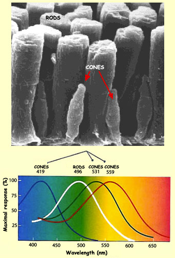
Our perception of color depends on the relative activity of three types of cones. Prevalence in retina 5-10. Learn vocabulary terms and more with flashcards games and other study tools.
Cones are active at higher light levels photopic vision are capable of color vision and are responsible for high spatial acuity. Rods are responsible for vision at low light levels scotopic vision. Main Differences between Rods and Cones Rods are responsible for vision in dim or night light while cones are responsible for vision in bright or daylight.
Less sensitive to dim light and less sensitive to fine detail. Both cells are packed with photoreceptive opsin proteins rhodopsin in rod and iodopsin in cone. Photopic daylight vision is controlled by the cones these cells require relatively bright light to function.
Cones help in colour vision. There are only three rods and three cones in each eye. Cones are active at higher light levels photopic vision are capable of color vision and are responsible for high spatial acuity.
The rods are more numerous some 120 million and are more sensitive than the cones. Response of rods is slow whereas that of cones is fast. Click card to see definition.
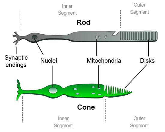
Cone-shaped photoreceptors found in the eye and are lesser in number compared to rods. Three types of cone cells that respond to different ranges of light wavelengths. Rod-shaped photoreceptors found in the eye imparting twilight vision.
More rods than cones in retina- about 100 million rod cells. They do not mediate color vision and have a low spatial acuity. Rods are the photoreceptor cells of the retina that are sensitive to dim light.
How many rods does each eye contain quizlet. Rods take long integration time while cones take short integration time. Photoreceptors are responsible for visual phototransduction.
How do rodscones influence y in the relation between the function of rodscones and their receptive fields respectively. They do not mediate color vision and have a low spatial acuity. The central fovea is populated exclusively by cones.
Do not impart color vision and are not differentiated. Compared with rods cones are_____a more sensitive to any light and less sensitive to fine detailb less sensitive to dim light and less sensitive to fine detailc more sensitive to dim light and less sensitive to fine detaild more sensitive to dim light and more sensitive to fine detaile less sensitive to dim light and more sensitive to fine detail. Rods and Cones are the photoreceptors found in the eye rods have rod-like structure and provide twilight vision while cones are of the cone shape fewer in number and provides the vision in the day or bright light.
According to the trichromatic theory of color vision. Main Difference Rods vs Cones. More sensitive to dim light and more sensitive to fine detail.
Cones have conical shapes while rods have a cylindrical shape. The 6 to 7 million cones provide the eyes color sensitivity and they are much more concentrated in the central yellow spot known as the. It can produce superior results for both sensitivity and acuity with rods having larger receptive fields compared to.
Less sensitive to dim light and more sensitive to fine detail. Cone-shaped mostly located at fovea respond to higher intensity light. Learn vocabulary terms and more with flashcards games and other study tools.
Rods are narrow and are present throughout the retina. Tap again to see term. The central fovea is populated exclusively by cones.
Start studying Comparison of Rods Cones. Rods help in twilight vision. Prevalence in retina 90-95.
Imparts color vision and can be differentiated into three types red blue and green. Compared with rods cones are. Start studying ap psych rods vs cones.
Rods are important for perception of light colors. Tap card to see definition. In humans rods cone and retinal ganglion cells are the 3 main photoreceptors.
Cones are the photoreceptor cells of the retina that are sensitive to bright light. They have visual violet pigment called as iodopsin.

How To See Averted Vision And Dark Adaptation Cosmic Pursuits Eye Facts Science Education Anatomy And Physiology

Eye Rods And Cones Illustration Image Search Results Eye Illustration Rods Image Illustration

Vision Anatomy And Physiology

The Retinal Neurons Are Classified Into Three Main Types Including Download Scientific Diagram

2 Sensations And Perception Eye Flashcards Quizlet

Psychology Unit 4 Part 1 Flashcards Quizlet

Pl7 Physiology Of Vision Diagram Quizlet

Neural Tunic Flashcards Quizlet

Chapter 2 Visual Psych Diagram Quizlet
Rods American Academy Of Ophthalmology

Exam 5 Retina Flashcards Quizlet
How Would You Compare And Contrast The Rod Cells And The Cone Cells In The Retina Of The Human Eye Socratic

Pin On Aia Course Human Vision

Rods Vs Cones Segmentation Physiology Cones

Pin On Brain

Lecture 15 Visual System Flashcards Quizlet
The Brain From Top To Bottom

How Would You Compare And Contrast The Rod Cells And The Cone Cells In The Retina Of The Human Eye Socratic
The Brain From Top To Bottom


Comments
Post a Comment